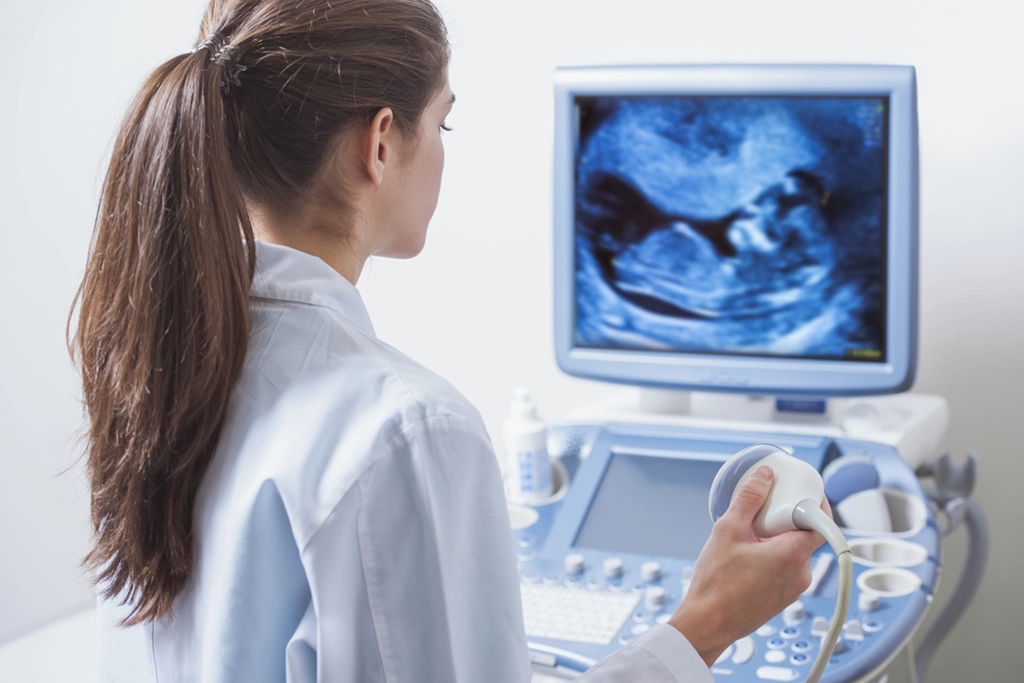
An ultrasound is a medical imaging technique that uses a transducer or probe, to generate sound waves and computer technology to produce pictures of the body's internal organs. Unlike other imaging techniques, an ultrasound creates “movies” that capture the movement of organs including heartbeats, contractions of bowel loops, and flow of blood vessels. An ultrasound is a safe and painless study where a small transducer gently follows a clear gel that is placed directly on the skin. The soundwaves admitted from the transducer pass through the gel, reflect off of the body, then are received by a computed to create an image. An ultrasound can evaluate different soft tissue structures in the body including the thyroid gland, breasts, abdomen, and pelvic organs.
Ultrasound


Our Ultrasound Locations
South Jersey Radiology offers ultrasound imaging services across 11 of our office locations. Each one of our office locations is conveniently located minutes away from major highways and bridges. With evening, weekend, and same-day appointments available, finding the answers you need has never been easier. View our office locations today.
Frequently Asked Questions
Understanding proper preparation and expectations can help alleviate any stress you may have prior to your appointment. Learn about what happens before, during, and after your ultrasound with our frequently asked questions.
When you arrive for your appointment, one of our ultrasound technologists will greet you and review the study with you. Afterward, you will be asked to lie down on a table where the area to be examined is exposed. A warm, clear gel will be applied to the area. Then, a small device called a transducer will pass over the gel and begin capturing images.
Once the study is complete, the gel will be wiped off and any portions that are not removed will dry to a powder. The gel will not stain or alter clothing.
An ultrasound requires very minimal preparation. Based on the specific area being studied, your doctor and our team of imaging experts at South Jersey Radiology will provide specific guidelines to follow before your study. Here are some general guidelines to follow:
- Arrive 15 minutes prior to your appointment.
- For an ultrasound of the liver, gallbladder, spleen, or pancreas, eat a low-fat dinner on the day before the exam and do not eat or drink for at least twelve hours prior to your appointment.
- For an ultrasound of your pelvis, you will need to have a full bladder. Please drink 6 eight-ounce glasses of water. Finish drinking this amount of liquid one hour prior to your appointment. Do not use the bathroom before your exam.
- For an ultrasound of your kidneys, you may be asked to drink 4 to 6 glasses of water one hour prior to your appointment.
- For an ultrasound of your aorta, please refrain from eating for at least 8 hours prior to your appointment.
An ultrasound is a painless and non-invasive imaging technique that uses no forms of radiation.
After your ultrasound appointment, one of our subspecialized radiologists will analyze the results and develop a detailed report for your doctor. Your doctor will receive the report within 48 hours and follow up with you to go over the results.
South Jersey Radiology is in-network with 99% of health insurance providers. Please contact your insurance provider to inquire about SJRA's in-network status. In some cases, insurance companies may attempt to tell you which radiology centers are preferred. As the patient, you have the right to choose if you would like your study performed at South Jersey Radiology.
